Background
Cancer therapy, such as chemo- and radiotherapy, aims to kill the cancer cells or stop their proliferative growth. Before being tested in animal models and applied in the clinic, all cancer therapies are first explored in vitro, i.e., in cell cultures in the laboratory, to understand the effects and mechanisms of action of the therapies in malignant and non-malignant cells, as well as to identify potential resistance mechanisms against the therapy that may exist or develop in the cells. Such preclinical exploration of cancer therapy is traditionally done in cells that are cultured in isolation (i.e., cultures consisting of cancer cells only or non-malignant cell only). Although useful, the knowledge obtained by this approach is limited in that it poorly mimics the therapeutic setting in cancer patients, whose tumours consist of not only cancer cells, but also of non-malignant cells that are intermixed and functionally engaged with the cancer cells. The mix of cancer cells and non-malignant cells that together make up the tumour is called “the tumour microenvironment”. It has recently been realized that cellular responses to cancer therapy can be very different if cancer cells and non-malignant cells of the tumour microenvironment are communicating, as compared to the responses of isolated cells to the same therapy. Therefore, front-line preclinical research on cancer therapy needs to include more advanced models than only isolated cell cultures. This Master project will employ co-culture models of cancer cells mixed with the most abundant non-malignant cell type in the tumour microenvironment (called “CAFs” – see below), to better understand the effects of clinically relevant cancer therapies in the tumour setting.
Intriguingly, cancer cells shape their own tumour microenvironment, in part by inducing changes in the normal cells that surround them. Particular attention has been given to the phenomenon that cancer cells can “reprogram” or differentiate surrounding fibroblasts to an activated state. In turn, these so-called “cancer-associated fibroblasts” (CAFs) promote tumour progression and may even mediate resistance to cancer therapy.
Research has suggested that tumour progression and therapy resistance are associated with alterations in the lysosomal recycling process called “autophagy”, and recent studies have indicated that autophagy may play a key role in the interplay between cancer cells and CAFs. However, this has thus far only been assessed by using autophagy markers, which are insufficient on their own to reliably monitor autophagic activity. Therefore, there is a need to employ functional autophagy assays in such investigations. Our laboratory is unique in that we have established and are mainly using - indeed - functional autophagy methods instead of markers to monitor autophagic activity, and this is leading to novel discoveries about the role of autophagy in cancer. Recently, we have found that co-culturing cancer cells and CAFs has a pronounced effect on autophagic responses to nutrient starvation. This warrants further investigation (=this Masters project) to explore whether and how autophagic responses to cancer therapy are altered by cancer cell-CAF co-culture, and whether such alterations in autophagy affect therapeutic efficacy.
CAFs are particularly abundant in the tumour microenvironment of solid tumours that arise from the malignant transformation of epithelial cells (“carcinomas”), e.g., in tumours of the colon, pancreas, lung, breast, and prostate. These 5 cancer types are the top 5 cancers with respect to the number of cancer-related deaths in Norway. Therefore, it is important to perform more research on the interplay between cancer cells and CAFs, and particularly in these cancer types. Prostate cancer is the most frequent of all cancer types and the third leading cause of cancer-related deaths in Norway, with ~5 000 new cases and ~1 000 deaths from prostate cancer in Norway each year. Besides surgery, the therapeutic options for prostate cancer patients with aggressive or advanced disease include hormone-, chemo-, and radiotherapy.
Aims and approaches
This project will employ co-cultures of cancer cells and CAFs derived from tumours of prostate cancer patients, and aims to:
- Investigate the abilities of clinically relevant prostate cancer therapies (hormone therapy, chemotherapy and radiation) to kill and/or slow down the proliferation of prostate cancer cells and CAFs when the cells are in co-culture compared to when the cells are in monoculture
- Employ unique functional autophagy assays to determine the effects of prostate cancer therapies on autophagy in prostate cancer cells and CAFs when the cells are in co-culture compared to when the cells are in monoculture
- Decipher the role of autophagy in the concerted responses of prostate cancer cells and CAFs to the cancer therapies by experimentally interfering with autophagy (knocking down autophagy-related genes) in each cell type and measuring the effects on cell proliferation and cell death. CRISPR-Cas knock-out cell lines may also be employed.
The prostate cancer cells are engineered to express red fluorescence, whilst the prostate CAFs express green fluorescence. This enables individual tracking of therapeutic effects as well as the possibility of sorting the cells by flow cytometry after co-culture for assessment of biological effects in the individual cell types. Figure 1 shows one example of such cells in physical co-culture:
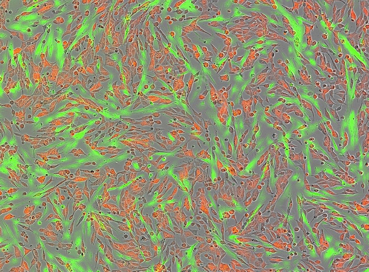
Figure 1. Physical co-culture of prostate CAFs (green-fluorescent, typical long and stretched cells) with prostate cancer cells (red-fluorescent, more roundish and less stretched cells). Dual-fluorescent image taken during live-cell imaging with an IncuCyte instrument.
Methods
- Sterile cell culture of prostate cancer cells and CAFs
- Setting up experimental co-cultures of prostate cancer cells and CAFs
- Experimental treatment of cells with hormone- and chemotherapy. Exposing cells to X-ray irradiation (similar to what is given to patients).
- Microscopy and live-cell fluorescence imaging
- Cell proliferation assays
- Cell death assays
- Flow cytometry
- siRNA transfection (knockdown) of autophagy-related genes
- Western blotting
- Cargo-based, functional autophagy assays. These include crude cell fractionation enabled by plasma membrane electrodisruption, biochemical assays, robotic measurements, and flow cytometry
Research environment and Expectations
You will be part of the “Autophagy in Cancer” Project Group (headed by Nikolai Engedal) - a supportive, focused, and passionate team with a variety of scientific backgrounds. You can expect an exciting Masters project in which you will learn scientific thinking and many useful techniques. You will also have the chance to make important novel discoveries at the forefront of the field of prostate cancer and autophagy research. There is scope for participating at a scientific conference. You can expect fully committed supervision and presence, and to take part in project group meetings, where you will get insights into other people’s work and can also get training in presenting your own work. We expect full time-commitment, scientific open-mindedness and curiosity, good communication skills, and a desire to work independently under guidance.
Contact
If you are interested in this project, please contact Nikolai Engedal nikolai.engedal@rr-research.no, Natalie Kurganovs Natalie.Jayne.Kurganovs@rr-research.no, or Cinzia Progida c.a.m.progida@ibv.uio.no.
We are happy to tell you more about the project and our research group, and to show you around at our facilities at the Department of Tumor Biology, Institute for Cancer Research, in the top modern Research Building at Radiumhospitalet (address: Ullernchausseen 70, 0379 Oslo).
Web site of the “Autophagy in Cancer” Project Group:
